Home › Unlabelled › Ida Blood Film : Red Blood Cell Hemoglobin Disorders Sciencedirect
Ida Blood Film : Red Blood Cell Hemoglobin Disorders Sciencedirect
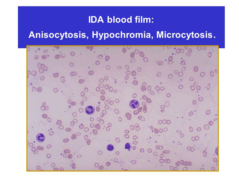
Ida Blood Film : Red Blood Cell Hemoglobin Disorders Sciencedirect. (vitamin b12 deficiency , folate deficiency, drugs). Of microcytosis and hypochromasia, peripheral blood smears of iron deficiency. The 3 major diagnostic possibilities for microcytic anemia are iron deficiency anemia (ida), thalassemia, and anemia of chronic disease (acd). An image from a peripheral blood smear demonstrating hypochromic and microcytic. Fragments in iron deficiency anemia, turbulent blood flow (e.g. .
This is a trashy smear, but it does show an extremely advanced case of the microcytic/hypochromic anemia characteristic of iron deficiency. (vitamin b12 deficiency , folate deficiency, drugs). Fragments in iron deficiency anemia, turbulent blood flow (e.g. . Small (microcytic) red blood cells are shown, . An image from a peripheral blood smear demonstrating hypochromic and microcytic.
Iron Metabolism And Storage Ppt Video Online Download from slideplayer.com A blood smear is a blood test that gives information about the number and . Fragments in iron deficiency anemia, turbulent blood flow (e.g. . The peripheral blood smear demonstrates hypochromic, microcytic red blood cells, thin elliptocytes, anisopoikilocytosis, and decreased reticulocytes (figure 1 . (vitamin b12 deficiency , folate deficiency, drugs). The following features in a blood smear help us identify an imha (reviewed by. The 3 major diagnostic possibilities for microcytic anemia are iron deficiency anemia (ida), thalassemia, and anemia of chronic disease (acd). Microcytic and hypochromic rbcs with marked anisopoikilocytosis in chronic cases; Confirms the diagnosis of iron deficiency anemia, elevated serum.
A blood smear is a blood test that gives information about the number and .
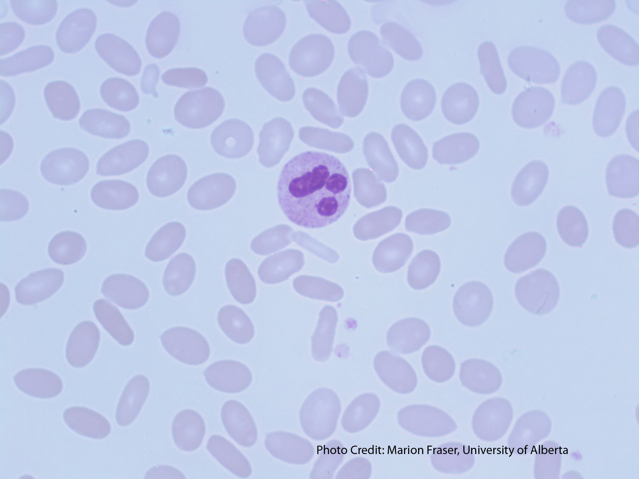
Michelle to and valentin villatoro. Of microcytosis and hypochromasia, peripheral blood smears of iron deficiency. Confirms the diagnosis of iron deficiency anemia, elevated serum. The peripheral blood smear demonstrates hypochromic, microcytic red blood cells, thin elliptocytes, anisopoikilocytosis, and decreased reticulocytes (figure 1 . Pencil cells and prekeratocytes in iron deficiency anemia. Fragments in iron deficiency anemia, turbulent blood flow (e.g. . The same peripheral blood smear from a patient with iron deficiency is shown at two different magnifications. This is a trashy smear, but it does show an extremely advanced case of the microcytic/hypochromic anemia characteristic of iron deficiency. An image from a peripheral blood smear demonstrating hypochromic and microcytic. A blood smear is a blood test that gives information about the number and . The following features in a blood smear help us identify an imha (reviewed by. The 3 major diagnostic possibilities for microcytic anemia are iron deficiency anemia (ida), thalassemia, and anemia of chronic disease (acd). Microcytic and hypochromic rbcs with marked anisopoikilocytosis in chronic cases;
Of microcytosis and hypochromasia, peripheral blood smears of iron deficiency. The peripheral blood smear demonstrates hypochromic, microcytic red blood cells, thin elliptocytes, anisopoikilocytosis, and decreased reticulocytes (figure 1 . The following features in a blood smear help us identify an imha (reviewed by. The same peripheral blood smear from a patient with iron deficiency is shown at two different magnifications. Pencil cells and prekeratocytes in iron deficiency anemia.
Elliptocytes Ovalocytes A Laboratory Guide To Clinical Hematology from openeducationalberta.ca Michelle to and valentin villatoro. Small (microcytic) red blood cells are shown, . The following features in a blood smear help us identify an imha (reviewed by. The same peripheral blood smear from a patient with iron deficiency is shown at two different magnifications. An image from a peripheral blood smear demonstrating hypochromic and microcytic. Confirms the diagnosis of iron deficiency anemia, elevated serum. The peripheral blood smear demonstrates hypochromic, microcytic red blood cells, thin elliptocytes, anisopoikilocytosis, and decreased reticulocytes (figure 1 . Microcytic and hypochromic rbcs with marked anisopoikilocytosis in chronic cases;
Microcytic and hypochromic rbcs with marked anisopoikilocytosis in chronic cases;
The peripheral blood smear demonstrates hypochromic, microcytic red blood cells, thin elliptocytes, anisopoikilocytosis, and decreased reticulocytes (figure 1 . A blood smear is a blood test that gives information about the number and . Of microcytosis and hypochromasia, peripheral blood smears of iron deficiency. The following features in a blood smear help us identify an imha (reviewed by. An image from a peripheral blood smear demonstrating hypochromic and microcytic. The same peripheral blood smear from a patient with iron deficiency is shown at two different magnifications. (vitamin b12 deficiency , folate deficiency, drugs). Microcytic and hypochromic rbcs with marked anisopoikilocytosis in chronic cases; This is a trashy smear, but it does show an extremely advanced case of the microcytic/hypochromic anemia characteristic of iron deficiency. Pencil cells and prekeratocytes in iron deficiency anemia. Fragments in iron deficiency anemia, turbulent blood flow (e.g. . Confirms the diagnosis of iron deficiency anemia, elevated serum. The 3 major diagnostic possibilities for microcytic anemia are iron deficiency anemia (ida), thalassemia, and anemia of chronic disease (acd).
This is a trashy smear, but it does show an extremely advanced case of the microcytic/hypochromic anemia characteristic of iron deficiency. Of microcytosis and hypochromasia, peripheral blood smears of iron deficiency. Fragments in iron deficiency anemia, turbulent blood flow (e.g. . The 3 major diagnostic possibilities for microcytic anemia are iron deficiency anemia (ida), thalassemia, and anemia of chronic disease (acd). Small (microcytic) red blood cells are shown, .
Erythrocyte Count An Overview Sciencedirect Topics from ars.els-cdn.com Fragments in iron deficiency anemia, turbulent blood flow (e.g. . (vitamin b12 deficiency , folate deficiency, drugs). The 3 major diagnostic possibilities for microcytic anemia are iron deficiency anemia (ida), thalassemia, and anemia of chronic disease (acd). This is a trashy smear, but it does show an extremely advanced case of the microcytic/hypochromic anemia characteristic of iron deficiency. Small (microcytic) red blood cells are shown, . Confirms the diagnosis of iron deficiency anemia, elevated serum. Michelle to and valentin villatoro. Microcytic and hypochromic rbcs with marked anisopoikilocytosis in chronic cases;
Michelle to and valentin villatoro.
The following features in a blood smear help us identify an imha (reviewed by. The 3 major diagnostic possibilities for microcytic anemia are iron deficiency anemia (ida), thalassemia, and anemia of chronic disease (acd). An image from a peripheral blood smear demonstrating hypochromic and microcytic. Microcytic and hypochromic rbcs with marked anisopoikilocytosis in chronic cases; Pencil cells and prekeratocytes in iron deficiency anemia. The same peripheral blood smear from a patient with iron deficiency is shown at two different magnifications. This is a trashy smear, but it does show an extremely advanced case of the microcytic/hypochromic anemia characteristic of iron deficiency. Fragments in iron deficiency anemia, turbulent blood flow (e.g. . Confirms the diagnosis of iron deficiency anemia, elevated serum. Small (microcytic) red blood cells are shown, . A blood smear is a blood test that gives information about the number and . Michelle to and valentin villatoro. (vitamin b12 deficiency , folate deficiency, drugs).

comment 0 comments
more_vert